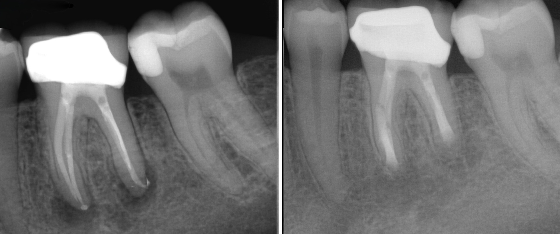
Periapical radiograph of tooth #30 showed a furcation radiolucency (infection). At 6-month follow-up, radiographs demonstrated complete healing with evidence of bone regeneration around the roots.
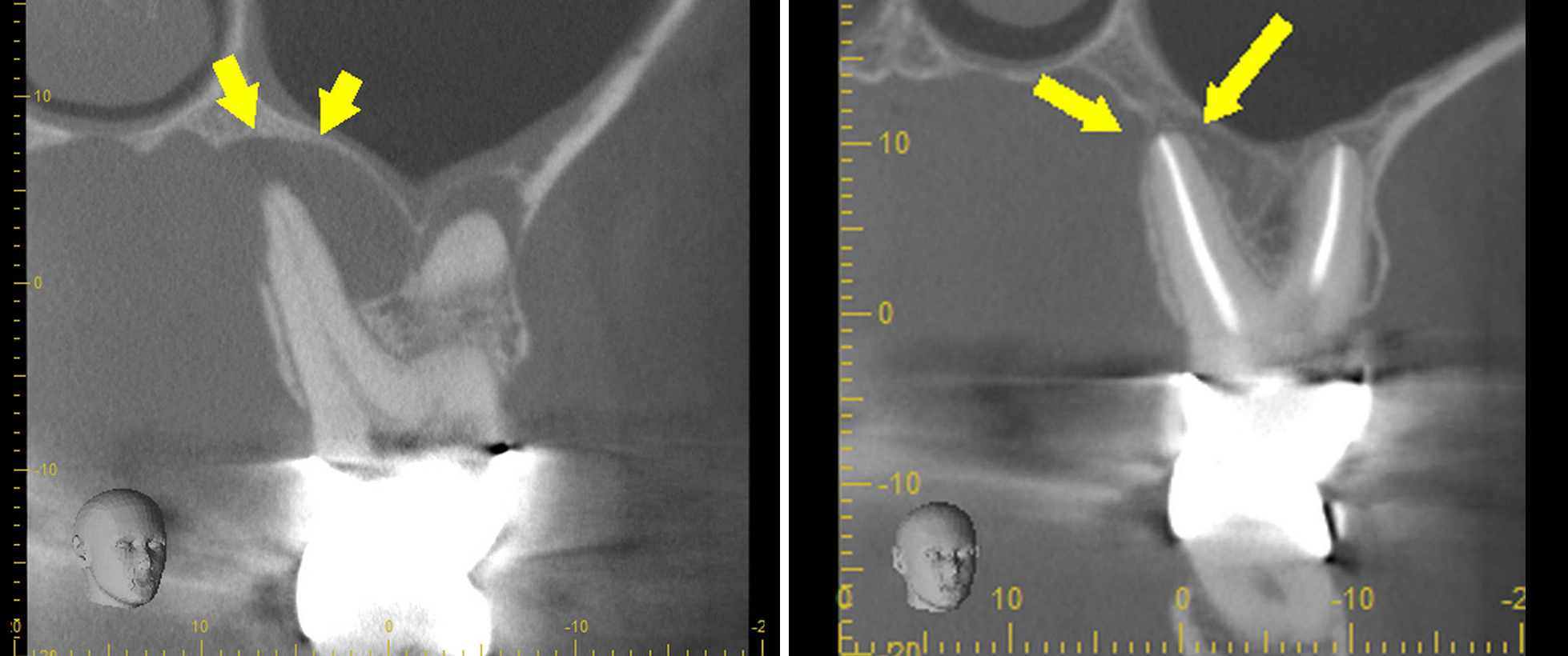
Sagittal CBCT view of tooth #14 reveals a large periapical radiolucency, most prominent around the palatal root, with close approximation to the floor of the maxillary sinus. Six-month follow-up imaging demonstrates significant osseous healing around all roots.
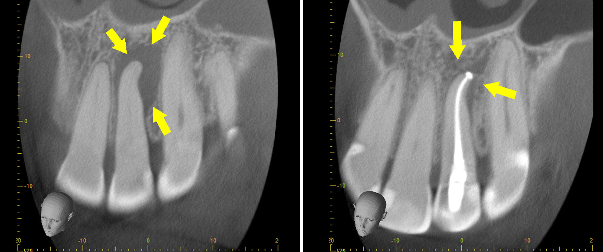
Coronal CBCT view of tooth #10 demonstrates a calcified pulp canal space and a large periapical radiolucency (infection) associated with the root apex. 6 month follow-up shows substantial periapical bone healing
Before-and-after X-rays of tooth #19 show the results of minimally invasive microsurgery. The infection around the root has cleared, and healthy bone has begun to regenerate around the previously treated tooth.
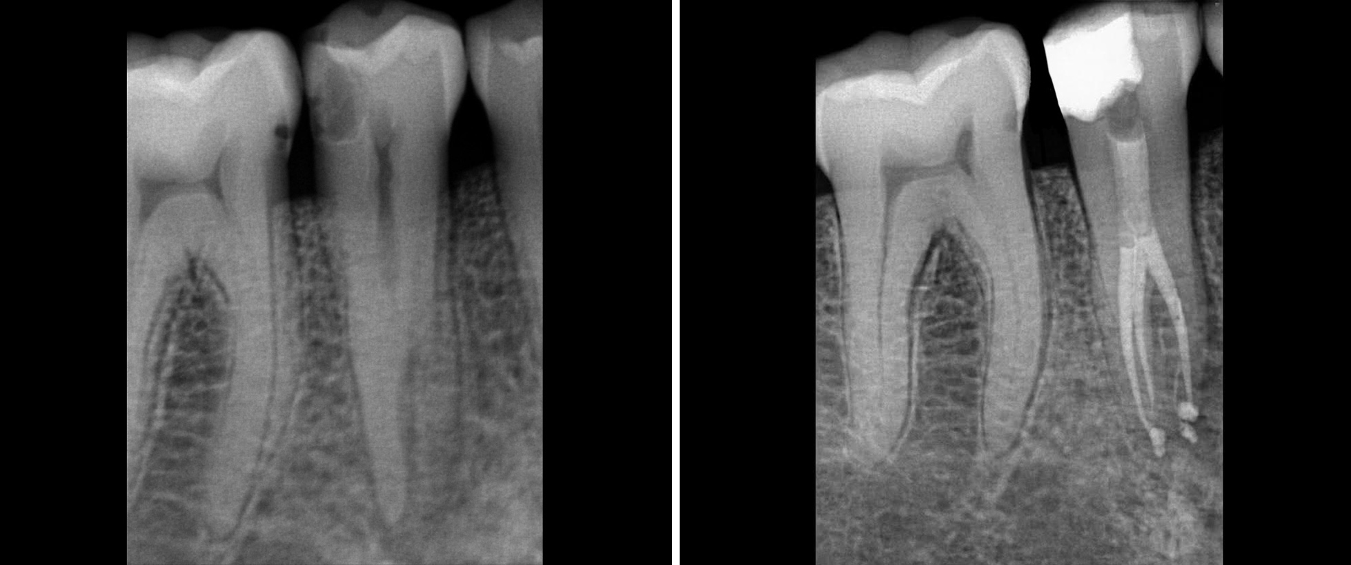
Pre- and post-op X-rays of tooth #30, the supporting tooth under a bridge, show severe calcification with no visible pulp space. While extraction was previously recommended, we successfully performed root canal treatment through the bridge—preserving the tooth and saving the patient’s new bridge.
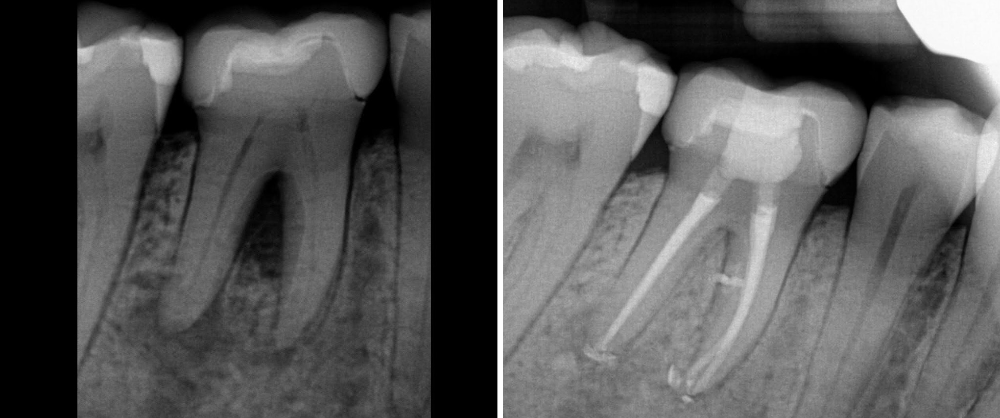
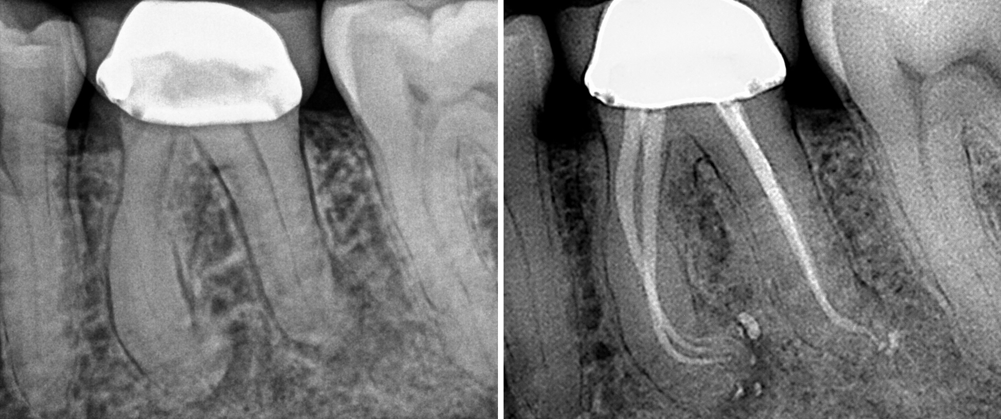
Pre- and post-op images of tooth #29. The patient presented with severe pain caused by deep decay extending to the nerve. Same-day treatment was performed to alleviate the pain. Post-operative radiograph shows a highly precise root canal fill despite complex anatomy—achieved through the integration of advanced technology and expert clinical skills.
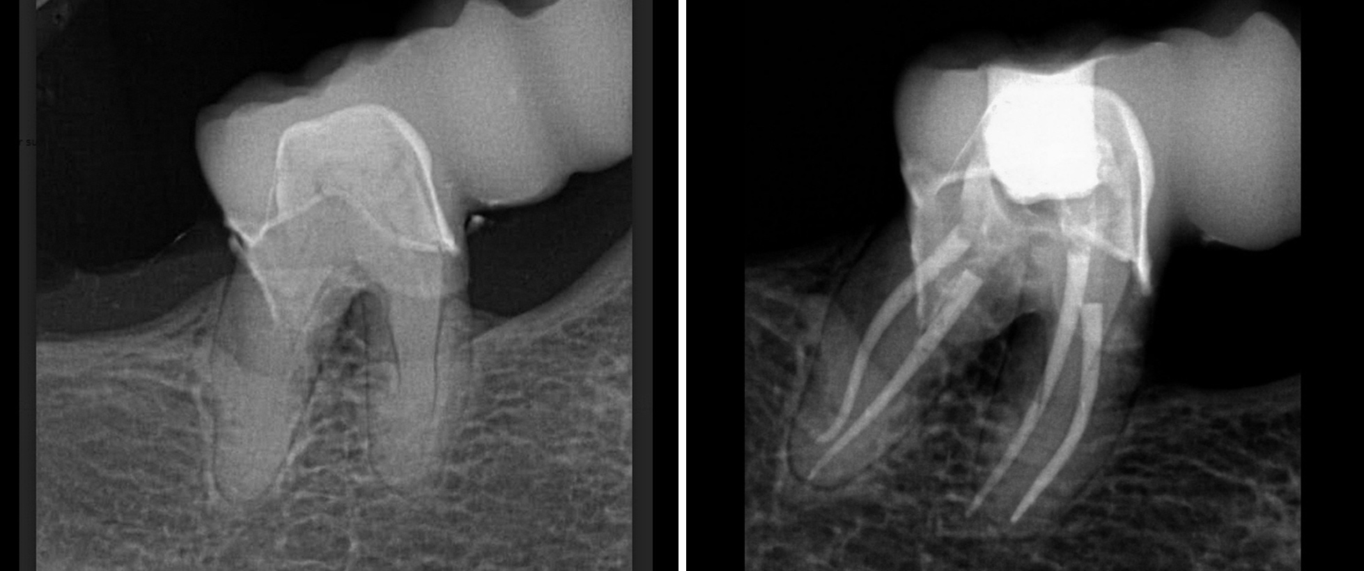
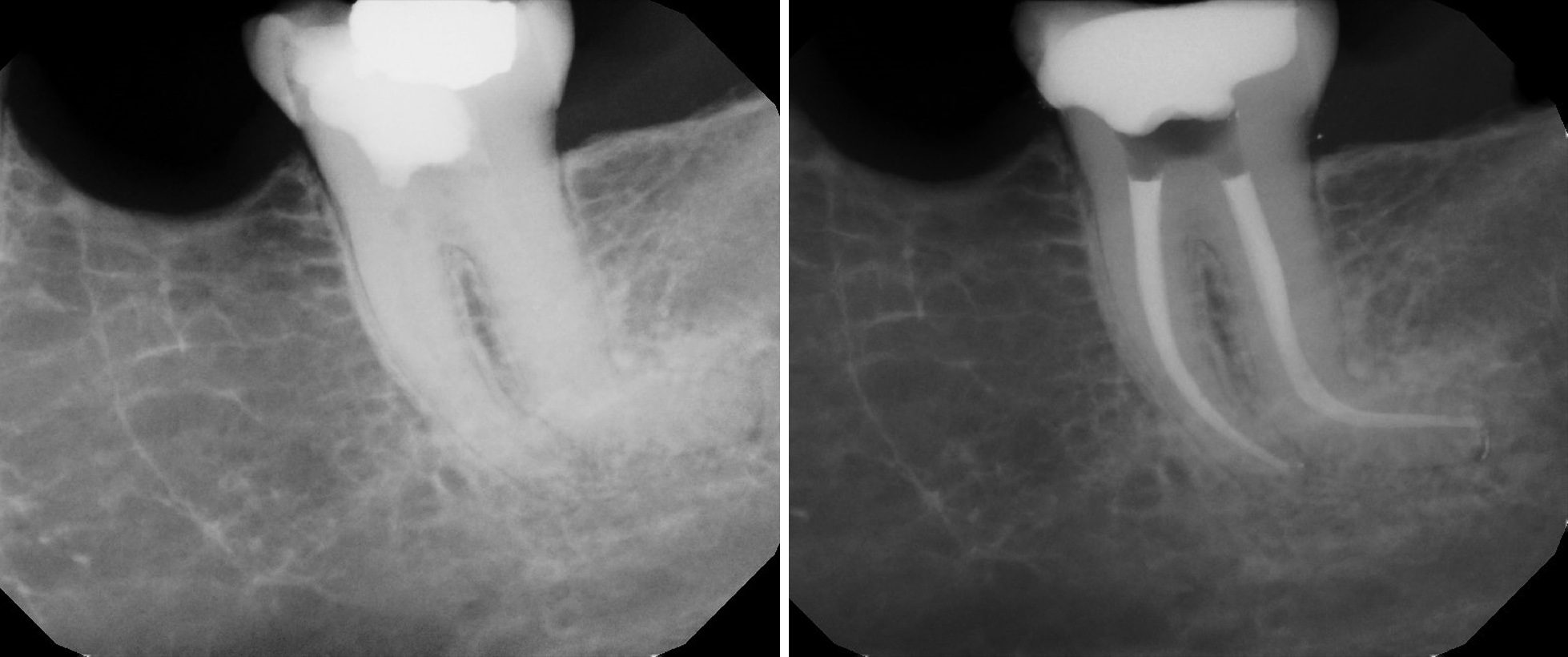
Pre- and post-op X-rays of tooth #19. The patient came in with excruciating pain. Despite the tooth’s complex root anatomy and severe curvature, we performed an emergency root canal through the crown. The final results show excellent treatment and pain relief.
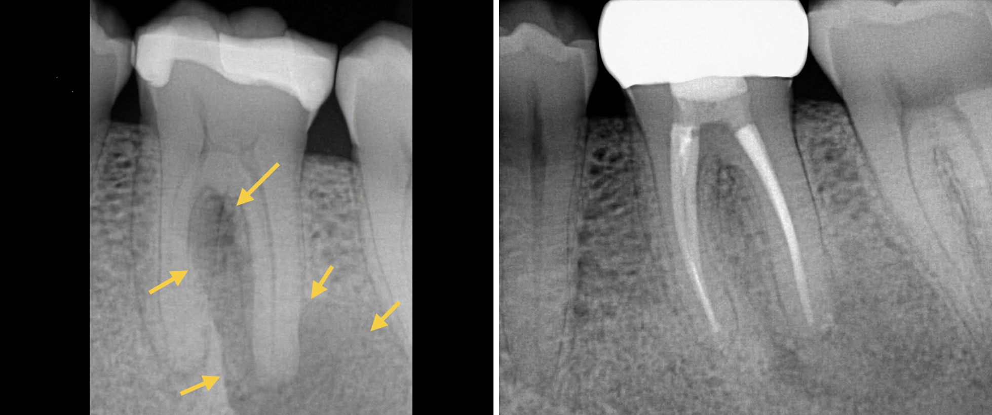
Pre- and post-op radiographs of tooth #19. The initial image shows a significant infection and and bone resorption (visible as a dark halo) around the root tip. Just six months after treatment, the follow-up X-ray shows excellent healing .
Pre- and post-op radiographs of tooth #18. This was a particularly challenging case due to a severe curvature in the distal root — a complexity even for experienced specialists. The post-op image shows a beautifully dense and precise root canal fill.
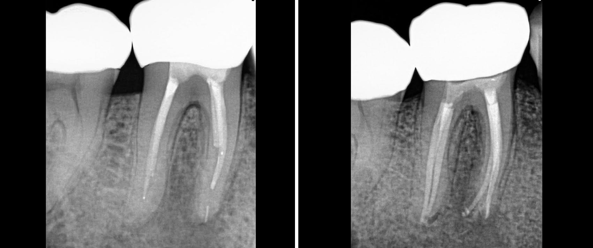
Pre-op radiograph of tooth #30 reveals a previously treated root canal with a separated instrument lodged at the bottom of the canal. The post-op image shows successful retreatment, with the instrument bypassed and the area thoroughly cleaned and disinfected.
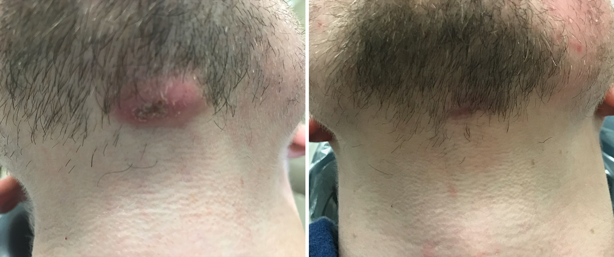
Pre-operative photo shows a severe extraoral infection originating from tooth #25. Remarkably, just two weeks after initiating root canal treatment, the post-op photo reveals complete healing of this extensive facial abscess.